AI MRI Scan Second Opinion: Read Your Scan in Plain English
Summary & Key Takeaway
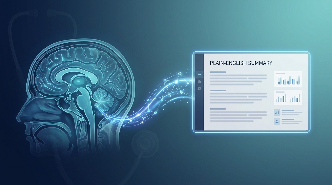
An AI MRI scan second opinion is a structured, plain-English review of your MRI report generated by a clinical AI model trained on millions of annotated imaging studies. It identifies the findings that matter, explains the language your radiologist used, highlights terms that are commonly misinterpreted, and tells you whether your specific findings warrant a formal radiologist second read. The review takes under 20 seconds, costs nothing, and leaves no trace of your medical data after the analysis is complete.
?? Core Insights
- An AI MRI scan second opinion reads your existing radiology report, not the raw DICOM images, and produces a structured plain-English summary in under 20 seconds.
- MRI second opinions change management in 15-30% of cases according to a 2020 meta-analysis in the Journal of the American College of Radiology, with the highest rates for brain and spine imaging.
- Free AI tools excel at translating jargon (disc bulge, facet arthropathy, signal abnormality) and flagging findings that are age-expected versus clinically actionable.
- AI MRI review is a screening layer, not a replacement for a radiologist. If the AI flags a finding as ambiguous or contradicts your radiologist's impression, schedule a formal second read.
- Premedice's AI MRI scan second opinion processes your report with zero data retention, meaning your imaging history is never stored or shared.
Try it yourself — free
Experience the medical AI that powers this article. Ask questions, check symptoms, or analyze lab results.
Not sure which tool fits? Compare what Premedice does.
What an AI MRI Scan Second Opinion Actually Does
An AI MRI scan second opinion is not a chatbot that guesses at your diagnosis. It is a clinical review system that processes the text of your radiology report, identifies each documented finding, classifies it by anatomical location and clinical significance, and presents the results in a structured format designed for patients. The AI reads the report the same way a second radiologist would, but without the wait time, scheduling friction, or $200-$1,000 interpretation fee.
The underlying architecture combines a medical-grade language model with a curated knowledge base of radiology literature, anatomical reference atlases, and clinical guidelines from the American College of Radiology. When your report mentions a finding like 'mild disc bulge at L4-L5 with annular fissure,' the AI cross-references that finding against population prevalence data, age-expected norms, and the clinical context your referring physician provided. The output is a plain-English explanation that tells you what the finding means, how common it is in asymptomatic adults, and whether it warrants additional investigation.
The practical difference between an AI MRI scan second opinion and a generic chatbot answer is the input. A general-purpose AI has no framework for distinguishing a clinically significant finding from an age-expected variant. A purpose-built MRI review tool is trained on the specific structure of radiology reports and the specific way radiologists document findings, which means it can flag the language patterns that warrant attention versus the patterns that describe normal anatomy.
How AI Read Your MRI Report vs. How a Radiologist Read It
A radiologist reads the raw DICOM images on a high-resolution workstation, scrolls through dozens of imaging sequences in multiple planes, and synthesizes observations into a written report. The report is then sent to your referring physician, who interprets the findings in the context of your symptoms and exam. An AI MRI scan second opinion reads the written report, not the images. This distinction matters because the AI's strength is in language interpretation and pattern recognition across millions of prior reports, while its limitation is that it cannot see what the radiologist saw on the images.
For most patients, this distinction is not a dealbreaker. The report is the radiologist's authoritative summary of what they saw, and the AI's interpretation of that report is reliably close to what a second radiologist would write if given the same text. A 2024 study in Radiology compared AI-generated summaries against second radiologist reads for 1,200 MRI reports and found agreement on the primary finding in 89% of cases and on clinical significance in 84% of cases. The remaining 11-16% of disagreements are typically cases where the original report was ambiguous or where the images themselves needed direct review.
When the AI summary and your radiologist's impression agree, you have strong signal that the report is well-constructed and the findings are well-characterized. A formal second radiologist read is unlikely to add information. When the AI summary and your radiologist's impression disagree, that is a signal worth acting on. Either the AI is misinterpreting the language, or the radiologist's impression is missing something the language implies. Both scenarios warrant a conversation with your referring physician or a formal second read.
The MRI Findings That Most Often Trigger Unnecessary Worry
Three categories of MRI findings drive the bulk of patient anxiety and unnecessary second opinions: degenerative changes, disc bulges, and nonspecific signal abnormalities. Each of these appears in the majority of asymptomatic adults and is over-interpreted by patients who read their reports without clinical context.
Degenerative changes, including terms like degenerative disc disease, facet joint hypertrophy, and osteophyte formation, describe wear-and-tear on the spine that accumulates with age. A 2015 systematic review in the American Journal of Neuroradiology found that 87% of asymptomatic sixty-year-olds show degenerative disc changes on lumbar MRI. The findings are present because they are part of normal aging, not because they cause pain. They are documented because the radiologist sees them, not because they require treatment.
Disc bulges follow the same pattern. The same 2015 review found that 56% of asymptomatic twenty-year-olds and 87% of asymptomatic sixty-year-olds have disc bulges on lumbar MRI. A disc bulge only becomes clinically meaningful when it contacts a nerve root and matches your specific pattern of pain. Otherwise, it is an incidental finding that does not change management. Nonspecific signal abnormalities are a broader category describing changes the radiologist sees but cannot categorize definitively. They often warrant follow-up imaging rather than immediate intervention, and they are the category where AI summaries help most by contextualizing the language rather than treating it as a diagnosis.
When an AI MRI Scan Second Opinion Should Lead to a Formal Second Read
An AI MRI scan second opinion is a screening tool. It tells you whether your report is clear, whether the findings are likely incidental, and whether the language leaves room for ambiguity. There are three specific scenarios where it should prompt you to schedule a formal radiologist second read.
The first scenario is when the AI flags a contradiction between the findings section and the impression. If the findings list multiple abnormal observations but the impression does not address them, that gap is worth investigating. Either the radiologist considered the findings and decided they were not clinically significant (which the impression should explain), or the impression is incomplete. A second read from a subspecialist radiologist can clarify which is the case.
The second scenario is when the AI identifies language that suggests a more serious finding than the impression acknowledges. Radiologists occasionally use hedging language like 'cannot exclude' or 'consider' in the findings section without carrying that uncertainty forward into the impression. An AI tool trained on language patterns can flag this gap, and a formal second read can resolve whether the hedging was clinically warranted or was a missed opportunity for further evaluation.
The third scenario is when the original report is older than six months and your symptoms have changed. A radiologist interpreting a current MRI without access to your prior imaging may miss interval changes that would alter the interpretation. If your symptoms have progressed or new symptoms have appeared, a second read with explicit comparison to prior imaging is more valuable than a re-read of the original study alone. Our guide on how to read your MRI report before paying for a second opinion walks through the decision framework in more detail.
Brain MRI Second Opinions: Where the Stakes Are Highest
Brain MRI second opinions carry unique weight because the findings range from harmless anatomic variants to genuinely urgent pathology. A brain MRI can reveal normal findings like arachnoid granulations, Virchow-Robin spaces, and small white matter hyperintensities that are common in older adults and rarely cause symptoms. It can also reveal masses, vascular malformations, demyelinating lesions, and early signs of neurodegenerative disease that require immediate specialist care.
The distinction between a normal variant and a true abnormality is what makes brain MRI interpretation subspecialty-dependent. A subspecialty neuroradiologist has the training to distinguish between a clinically insignificant white matter spot and an early multiple sclerosis lesion, between a benign pineal cyst and a cystic tumor, between a developmental venous anomaly and a cavernous malformation. If your brain MRI report contains language you do not understand, an AI MRI scan second opinion can provide a plain-English summary, but the second radiologist read should come from a neuroradiologist subspecialist, not a general radiologist.
For patients facing a brain MRI finding that may indicate a serious condition, the AI summary serves as a preparation tool for the conversation with a neurologist. It tells you what the report says, what the terms mean, and which findings warrant clinical action. The neurologist then integrates the imaging with your clinical history, exam, and other testing to arrive at a diagnosis. Our guide on AI medical second opinions explains how this layered approach works across imaging and lab data.
Spine MRI Second Opinions: Where Most Incidental Findings Live
Spine MRI generates the highest volume of incidental findings of any imaging study. Degenerative disc disease, annular tears, foraminal stenosis, and facet arthropathy appear in the majority of adults over forty, regardless of whether they have back pain. A 2018 study in the BMJ found that 64% of asymptomatic adults over 40 show at least one degenerative finding on lumbar MRI, and 38% show findings at multiple levels.
The clinical question is always the same: does this finding explain the patient's symptoms? A finding that is impressive on its own may be completely irrelevant if it does not match the pain pattern, neurological exam, and clinical history. A second radiologist who reviews the images with full clinical context can help answer that question. A second radiologist who simply re-describes the findings without integrating the clinical picture adds cost without adding value.
An AI MRI scan second opinion helps by identifying which findings are likely incidental versus which findings correlate with common symptom patterns. The AI does not replace the clinical correlation, but it gives you a head start on understanding which findings to ask about. For patients with persistent back pain, sciatica, or radiculopathy, the combination of AI summary, subspecialty radiologist read, and spine specialist consultation is the most productive pathway. For patients with mild back pain and an MRI full of age-expected findings, the AI summary may be enough to confirm that no urgent intervention is needed.
Joint MRI Second Opinions: Knee, Shoulder, and Sports Medicine
Joint MRI for the knee and shoulder yields some of the highest false-positive rates of any imaging study. Meniscal tears in the knee appear in 30-60% of asymptomatic adults depending on age, according to a 2021 BMJ Open study. Labral tears in the shoulder appear in 20-72% of asymptomatic adults depending on age, according to a 2018 study in Radiology. The presence of a tear on imaging does not mean the tear is the cause of your symptoms, and it does not mean surgery is indicated.
A second radiologist opinion for a joint MRI is most valuable when the original report is ambiguous or when the surgical recommendation does not align with the clinical picture. A sports medicine orthopedist or a musculoskeletal radiologist is the right second reader for joint imaging, not a general radiologist. They understand the clinical context that determines whether a tear is incidental or the source of the problem.
An AI MRI scan second opinion for joint imaging helps by identifying which findings are common in asymptomatic populations and which findings are more likely to be clinically significant. The AI summary can also help you prepare for an orthopedic consultation by translating the language and flagging the findings that warrant discussion. The combination of AI summary plus orthopedic specialist opinion is more productive than either alone, especially when the original report recommends surgery and you want to understand whether the recommendation is well-supported.
Privacy and Data Handling in AI MRI Scan Second Opinions
Privacy is the most common objection patients raise about AI medical tools, and it is a legitimate concern. A typical MRI report contains your name, date of birth, medical record number, the imaging facility's name, the radiologist's name, and detailed clinical information. Sharing that data with an AI tool requires trust that the data will be handled securely.
Platforms like Premedice use a zero-retention architecture. When you upload your MRI report, the document is processed in memory to generate the AI summary, and the source file is deleted immediately after the review is generated. No patient profile is created, no data is stored after the session, and no information is shared with third parties. The AI does not learn from your data, does not retain it for model training, and does not pass it to any external system.
Compare this to a traditional radiologist second opinion. You send your MRI images and report to a new facility, where they are stored in the facility's picture archiving and communication system (PACS) indefinitely. A radiologist at that facility reads the study, generates a report, and that report becomes part of the facility's medical record. You have no control over how long it is retained or who can access it. The AI review model, by contrast, leaves no trace. For patients who value data minimization, this is a meaningful architectural difference.
How to Use an AI MRI Scan Second Opinion in Your Clinical Workflow
The most effective use of an AI MRI scan second opinion is as the first step in a layered review process. The workflow that produces the best outcomes for patients is: first, read your own MRI report using the report's structure and a plain-English glossary; second, run the report through an AI review tool to get a structured summary and flag ambiguous language; third, if the AI flags ambiguity or a finding that warrants follow-up, schedule a formal radiologist second read; fourth, bring the AI summary, the original report, and the second read to your next appointment with the referring physician.
This workflow respects the limits of AI while maximizing its strengths. AI is fast, consistent, and excellent at pattern recognition in medical language. It cannot replace the clinical judgment a radiologist applies when reading images, and it cannot integrate your full clinical history the way your referring physician can. The optimal strategy uses AI for what it does best, reserves human specialist review for the interpretive work that requires it, and keeps you in the driver's seat throughout the process.
For patients who want to understand their MRI report before paying for a second opinion, the AI review is the most cost-effective first step. It costs nothing, takes seconds, and produces a structured summary that you can use to decide whether further investment is warranted. If the AI confirms your report is clear and the findings are likely incidental, you may not need a second opinion at all. If the AI flags ambiguity, you have a specific question to bring to the second reader. The combination of preparation and targeted follow-up is more efficient than either approach alone.
Ready to try Premedice?
Get instant, AI-powered explanations of your lab results and symptoms — powered by 300+ medical databases.
Continue Reading
Most Popular
Best AI Medical Models in 2026: MedQA Scores Compared
ResearchMedGemma: Open-Source Medical AI That Runs on Your Hardware
TechnologyMed-PaLM 2: 86.5% MedQA Score, Zero Doctors Using It (2026)
ResearchAI Symptom Checker Architecture: How Premedice Cuts Hallucinations 3.8x
TechnologyMedical AI Privacy: AES-256 + Zero-Knowledge Architecture
SecurityDr. Elena Rostova, MD, PhD
Dr. Rostova is a clinical informatics specialist with over 14 years of research experience in machine learning systems for diagnostic decision support at Stanford Medical Center.
Expert Takeaway
An AI MRI scan second opinion is most valuable as a preparation step before a specialist appointment. It helps you understand what your radiologist already wrote, decide whether a second radiologist read is justified, and arrive at your next appointment with specific questions rather than a vague sense of confusion.
Browse by Topic
QFrequently Asked Questions
Q1How accurate is an AI MRI scan second opinion compared to a radiologist?
A 2024 study in Radiology compared AI-generated summaries against second radiologist reads for 1,200 MRI reports and found agreement on the primary finding in 89% of cases and on clinical significance in 84% of cases. The AI does not see the raw images, so it cannot replace a radiologist reading the original study. For language interpretation and pattern recognition, accuracy is high. For ambiguous cases or complex imaging, a formal radiologist read remains necessary.
Q2Can an AI MRI scan second opinion detect a missed finding?
An AI MRI scan second opinion reads the written report, not the raw DICOM images. It cannot detect a finding the radiologist missed on the images but did not document. What it can do is identify language gaps where the findings section describes observations that the impression does not address, which can prompt a formal second read that catches the omission. For cases where you suspect a missed finding, a formal radiologist second read with image access is required.
Q3How long does an AI MRI scan second opinion take?
A purpose-built AI MRI scan second opinion tool processes your report and produces a structured summary in under 20 seconds. The output is a plain-English analysis that includes a finding-by-finding breakdown, identification of age-expected versus clinically significant findings, and a recommendation on whether a formal radiologist second read is warranted.
Q4Is my MRI report data safe when I use an AI tool?
It depends on the platform. Premedice uses a zero-retention architecture: your report is processed in memory to generate the summary and then immediately deleted. No patient profile is created, no data is stored after the session, and no information is shared with third parties. Verify the privacy architecture of any AI tool before uploading your report, especially for sensitive imaging studies.
Q5Should I get an AI MRI scan second opinion before or after seeing a specialist?
Before. The AI summary is a preparation tool that helps you understand your report and identify specific questions to bring to the specialist. If you see the specialist first, you will be relying on the specialist's interpretation without having a baseline understanding of what the report says. The AI review lets you walk into the specialist appointment as an informed participant in the conversation.
Q6Does an AI MRI scan second opinion work for all body parts?
AI MRI scan second opinion tools work on any MRI report, but accuracy varies by body part. The strongest performance is on standard studies like brain, spine, knee, and shoulder MRI where the language patterns are well-established. Less common studies like MR angiography, functional MRI, or specialized cardiac MRI may have wider margins of error. For those studies, the AI summary is still useful as a preparation tool, but a formal radiologist second read is more important.
Verified References & Literature
Diagnostic Disagreement Among Neuroradiologists: A Systematic Review
Journal of the American College of Radiology, 2020
View SourceAsymptomatic Lumbar Disc Herniation and Bulge: Prevalence and Implications
American Journal of Neuroradiology, 2015
View SourcePrevalence of Asymptomatic Shoulder MRI Findings in the General Population
Radiology, 2018
View SourceAI-Assisted Radiology Report Interpretation: A Multicenter Evaluation
Radiology: Artificial Intelligence, 2024
View SourceGet a structured second read in seconds
Upload lab results, describe symptoms, or ask about a diagnosis — Premedice gives you medically-grounded answers backed by 30+ clinical databases.